
Research Journey
What we love about iGEM is that it values not only results, but the effort, creativity, and persistence behind every step of the scientific journey. It reminds us that even the smallest contribution to science matters, especially when it comes from young researchers who are still learning how the world of synthetic and molecular biology works. We understood from the beginning that conducting advanced research within a few months is highly ambitious for any high-school team. In many regions, including ours, this challenge is multiplied by the limited access to laboratories, lack of funding, and the general unfamiliarity of high-school research programs among scientists.
When we first started this project, our goal was to go beyond in-silico design and develop a complete experimental system. We aimed to construct the biosensor plasmid through Golden Gate Assembly, transfect it into mammalian cells, and analyse the reporter response experimentally. Our original plan included observing EGFP expression through fluorescence microscopy to confirm successful transfection and activation of the ARE-driven reporter, alongside Western blot analysis of our key oxidative-stress proteins NRF2 for deep analysis.
However, as a high-school team working with limited funding, time, and access to reagents, we faced significant challenges in obtaining certain antibodies and materials. In particular, NRF2 antibodies were unavailable or prohibitively expensive, and delays in shipping prevented us from accessing many specialized reagents before our experimental deadline.
Given these constraints, we decided to focus on feasible, resource-based validation methods. The HSP70 antibody was available to us, and because hydroquinone exposure is known to trigger both oxidative and proteotoxic stress, we used HSP70 as an orthogonal marker to confirm that our treatment induced a general cellular stress response. Although this approach does not directly measure Nrf2 activation, it allows us to demonstrate the biological relevance of our system and support the stress conditions under which our reporter construct would normally be activated.
In essence, our experiments serve as a proof of concept - we designed and constructed the plasmid both in silico through Golden Gate Assembly. This establishes the foundation for the next stage of our project. In the coming iGEM, we aim to transfect HEK293T cells, visualize EGFP expression through fluorescence microscopy, and perform Western blot analysis with pathway-specific antibodies to fully validate the Nrf2-ARE biosensor system.
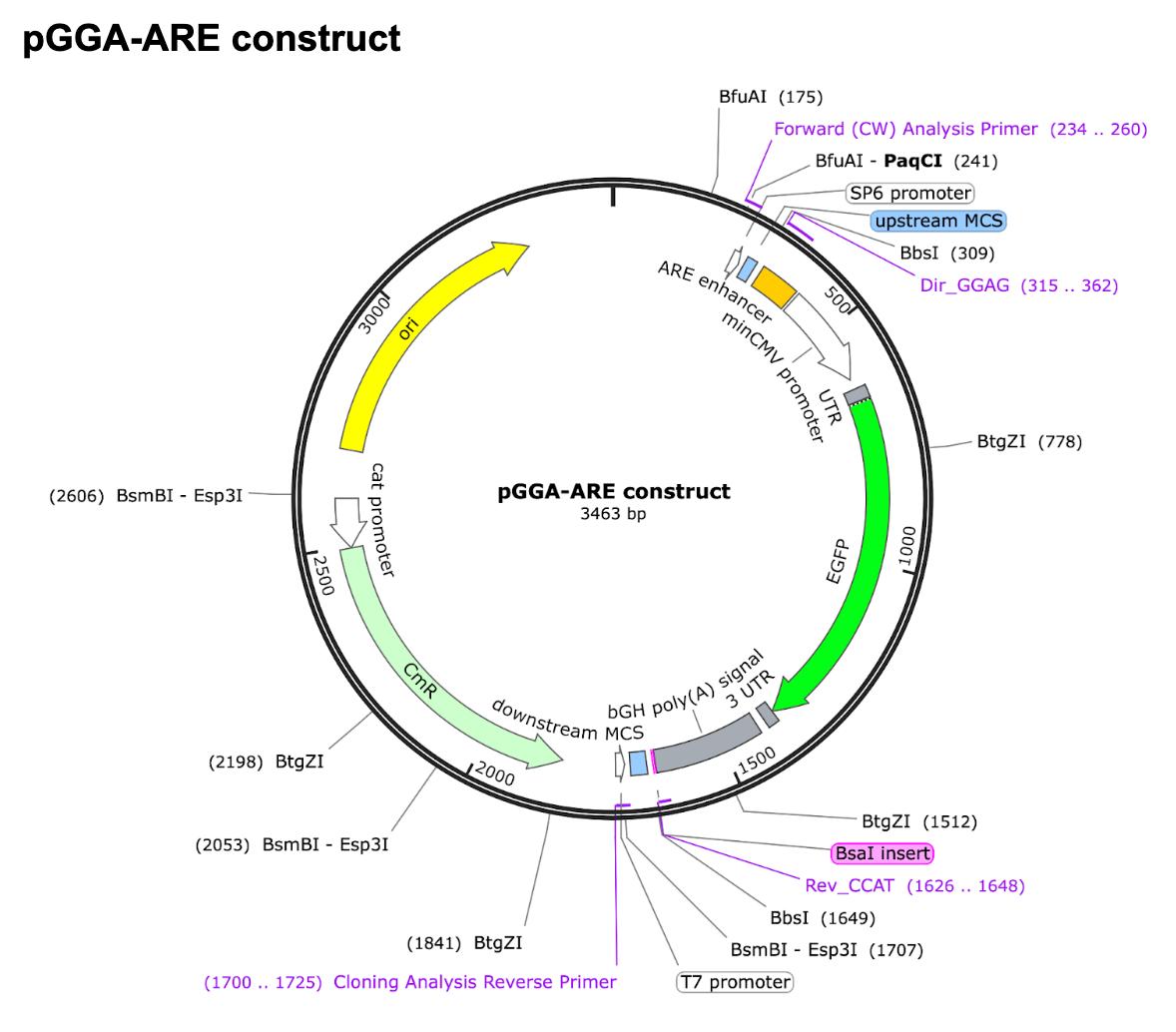
pGGA-ARE construct
In our experimental plan, we used the basic parts provided by the Asimov Mammalian parts. Specifically, we build our construction using the following parts: BBa_J433000, BBa_J433045, BBa_J433045, BBa_J433021, BBa_J428076 and added our new basic part: BBa_2508GVGJ.
We changed the full CMV enhancer- CMV promoter to minimal CMV promoter by removing its enhancer region. We then added the ARE enhance upstream of the minCMV promoter. We added the regulatory components such as 5' UTR, Kozak sequence, EGFP, 3' UTR, and bGH Poly(A) signal.
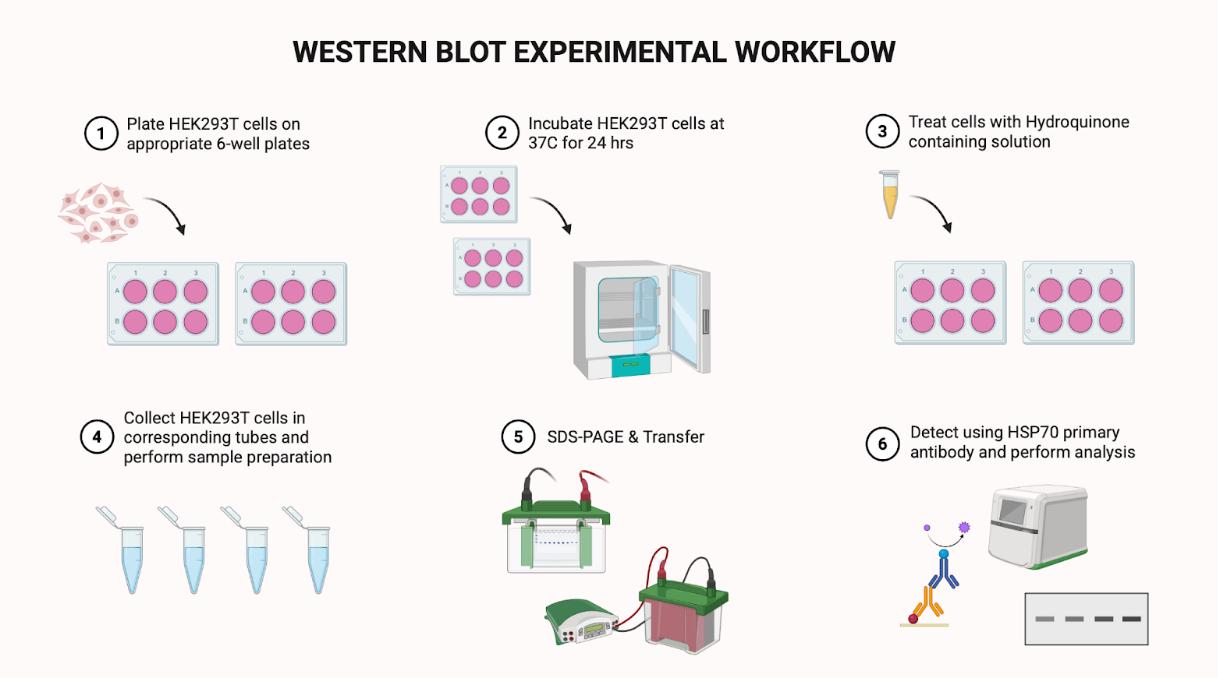
Western Blot
Our original plan was to analyze the Nrf2 protein, but because we did not have the specific antibody, we adapted our method and chose to detect HSP70, another well-known protein upregulated under stress conditions.
We prepared our cell lysates, quantified the protein concentrations, and loaded them onto SDS-PAGE gels. After transferring the proteins onto membranes, we performed immunoblotting using the HSP70 antibody.
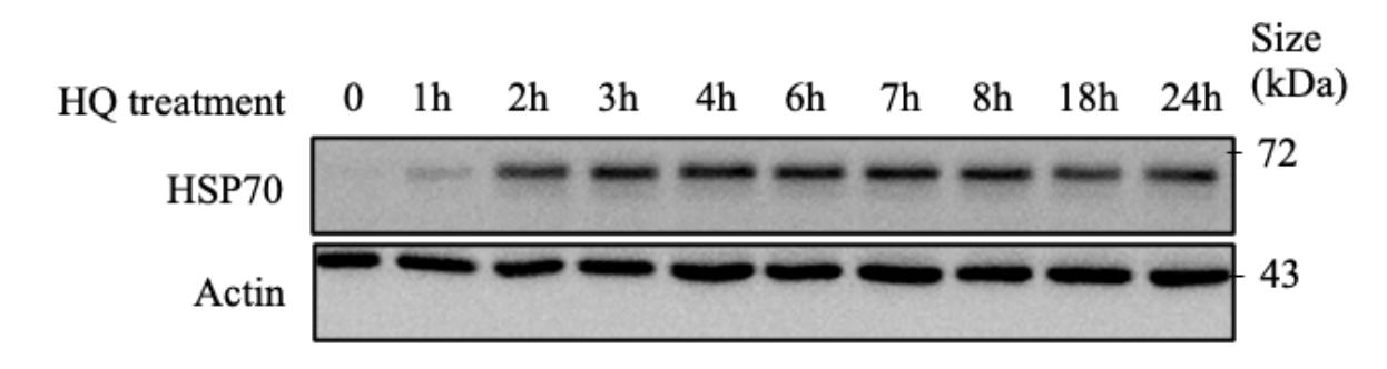
As you can see from the diagram above, we did not observe an immediate increase in HSP70 protein levels following the initial exposure to the hydroquinone-containing solution. However, after one hour, faint protein bands began to appear, suggesting the early activation of the stress response pathway. This indicates that hydroquinone activates a gradual cellular adaptation rather than an immediate reaction. After two hours, we detected a clear increase in HSP70 expression, which remained consistently elevated up to eight hours of exposure. This prolonged expression pattern illustrates that cells continuously respond to ongoing oxidative or proteotoxic stress induced by hydroquinone. After 18 hours, we observed a slight decline in HSP70 levels. We think this is reflecting cellular exhaustion or the natural attenuation of the stress response after long hours of stimulation. We used actin as the loading control to confirm equal protein loading across samples to ensure that the observed changes represent true variations in HSP70 expression.
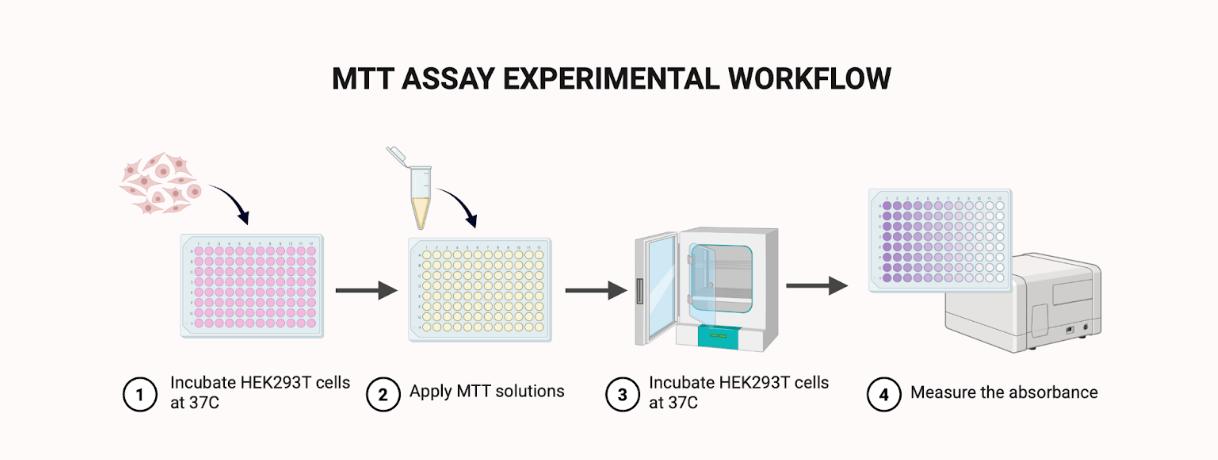
MTT Assay/h2>
Another additional experiment we conducted was the MTT assay. We used this experiment to test whether our HEK293T cells could respond to oxidative stress caused by hydroquinone (HQ), since we couldn't yet test our ARE-minCMV-EGFP plasmid in the lab. The MTT test shows how many cells are alive and active after being exposed to a chemical. Living cells can turn the yellow MTT solution into a purple color, and we measure that color at 570 nm using a plate reader. The darker the purple color, the higher the absorbance, which means more living cells.
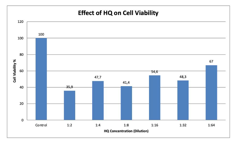
In our experiment, the control group (cells without hydroquinone) showed the highest absorbance value, meaning the cells were healthy. When we added hydroquinone, the absorbance dropped depending on how strong the concentration was. At higher concentrations, the absorbance was much lower, showing that more cells were damaged/dead. As we diluted the hydroquinone, the absorbance increased again, meaning more cells survived. This result shows that hydroquinone is toxic to cells and that our HEK293T cells can sense and respond to changes in their environment. Even though we couldn't test our biosensor plasmid yet, this MTT experiment confirmed that the cells react to oxidative stress, the same signal our plasmid is designed to detect by expressing the EGFP.