
Project Overview
We aim to develop a HEK293T cell-based biosensor or bio-reporter capable of detecting the presence of hydroquinone, a harmful and often illegally used compound in cosmetic products. Our long-term vision is to create an easy-to-use, affordable, and portable detection system, as simple as a pregnancy test, to help consumers identify counterfeit or unsafe cosmetics that contain this banned ingredient.
This project represents our proof-of-concept stage. We want to demonstrate that cells can act as sensitive, ethical biosensors for harmful cosmetic ingredients. Our long-term goal is to support public health, consumer safety, and regulatory monitoring while offering a non-animal alternative to current testing methods.
The experimental aspects of our project include:
- Chassis Selection
- Hydroquinone Metabolism & Mechanism of Action
- The Keap1-Nrf2 pathway
- ARE-minCMV-EGFP Construct For a Bio-sensor
Chassis Selection
We selected the HEK293T cell line as the chassis for our biosensor due to its numerous advantages in mammalian synthetic biology. These cells are easy to culture and maintain, capable of rapid proliferation, and can be grown in serum-free suspension systems if needed. Most importantly, HEK293T cells are highly amenable to transfection using reagents such as Lipofectamine, enabling efficient delivery and expression of plasmid DNA constructs.
Hydroquinone Metabolism & Mechanism of Action
Hydroquinone (HQ; 1,4-dihydroxybenzene) is an aromatic compound structurally similar to melanin precursors such as L-DOPA and dopamine (Figure 1). Because of these similarities, hydroquinone can enter the melatonin producing pathway and act as a substrate for tyrosinase, the copper-containing enzyme responsible for catalyzing the oxidation of phenolic compounds during melanin biosynthesis. This interaction marks the beginning of hydroquinone's metabolism in melanocytes and underlies both its depigmenting properties and its cytotoxic potential.
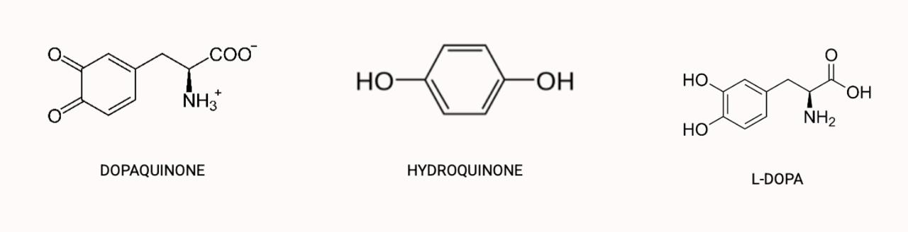
Figure 1. The structure of hydroquinone, dopaquinone and L-DOPA
At the biochemical level, hydroquinone interferes with melanin formation in three major ways:
- It competitively inhibits tyrosinase, reducing the conversion of L-DOPA to dopaquinone and thereby suppressing melanin synthesis.
- The oxidative conversion of hydroquinone to p-benzoquinone produces Reactive Oxygen Species (ROS) such as superoxide anion (O₂•⁻) and hydrogen peroxide (H₂O₂). These directly damage melanosomal components, including tyrosinase itself.
- Continuous oxidative and electrophilic stress leads to melanocyte degeneration, resulting in depigmentation.
These mechanisms explain why hydroquinone is widely used in skin-lightening formulations. Figure 2a and 2b demonstrate the schematic pathway of hydroquinone's action during the melanin synthesis and the potential result, respectively.
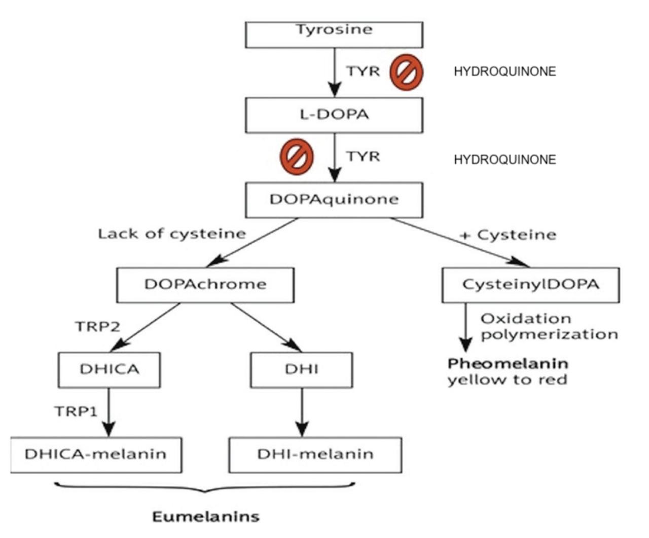
Figure 2a. The diagram of Hydroquinone's action during the melanin synthesis
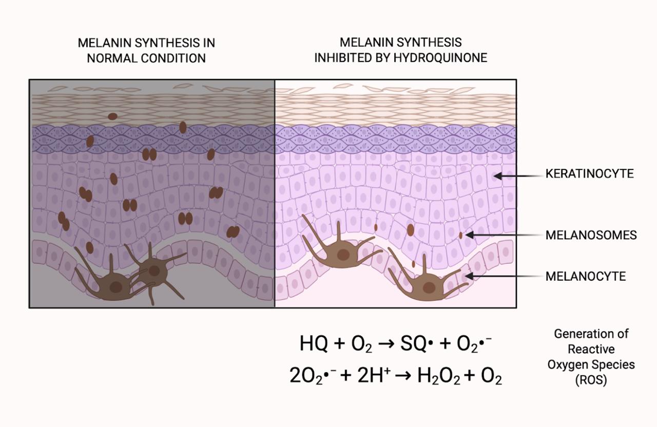
Figure 2b. The visual differences between the normal and hydroquinone applied skin conditions. The reaction shows the produced ROS inside these cells
The Keap1-Nrf2 Pathway
Our cells are constantly exposed to various stresses, including UV radiation, toxins, chemicals, and drugs. In our case, hydroquinone serves as a key example. The generation of reactive oxygen species (ROS) such as superoxide anion (O₂•⁻), hydrogen peroxide (H₂O₂), and hydroxyl radical (•OH) leads to an imbalance between oxidant production and antioxidant defenses. Under physiological conditions, ROS play essential signaling roles in cell proliferation, differentiation, and immune defense. However, when produced excessively, as occurs during the oxidation of hydroquinone, this can cause oxidative stress.
Yet, nature has evolved beautiful mechanisms to protect itself and maintain balance within cells, preventing permanent damage or even cell death. To counteract the cytotoxic effects of hydroquinone metabolism, cells can activate defense mechanisms. During oxidative stress, they initiate one of the major signaling cascades designed to restore internal homeostasis.
The same ROS that causes oxidative stress also acts as signaling molecules. Inside the cell, a specialized regulatory system known as the Keap1–Nrf2 pathway monitors and controls this process. ROS are sensed by redox-sensitive proteins, particularly those containing cysteine residues, which undergo reversible modifications in response to oxidative changes. Keap1 (Kelch-like ECH-associated protein 1) is one such protein and plays a central role in regulating the transcription factor Nrf2 (Nuclear factor erythroid 2-related factor 2).
Under normal conditions, Keap1 binds Nrf2 in the cytoplasm and leads to its ubiquitination and proteasomal degradation by Rbx1 and Cul3 proteins, keeping Nrf2 levels low (Figure 3).
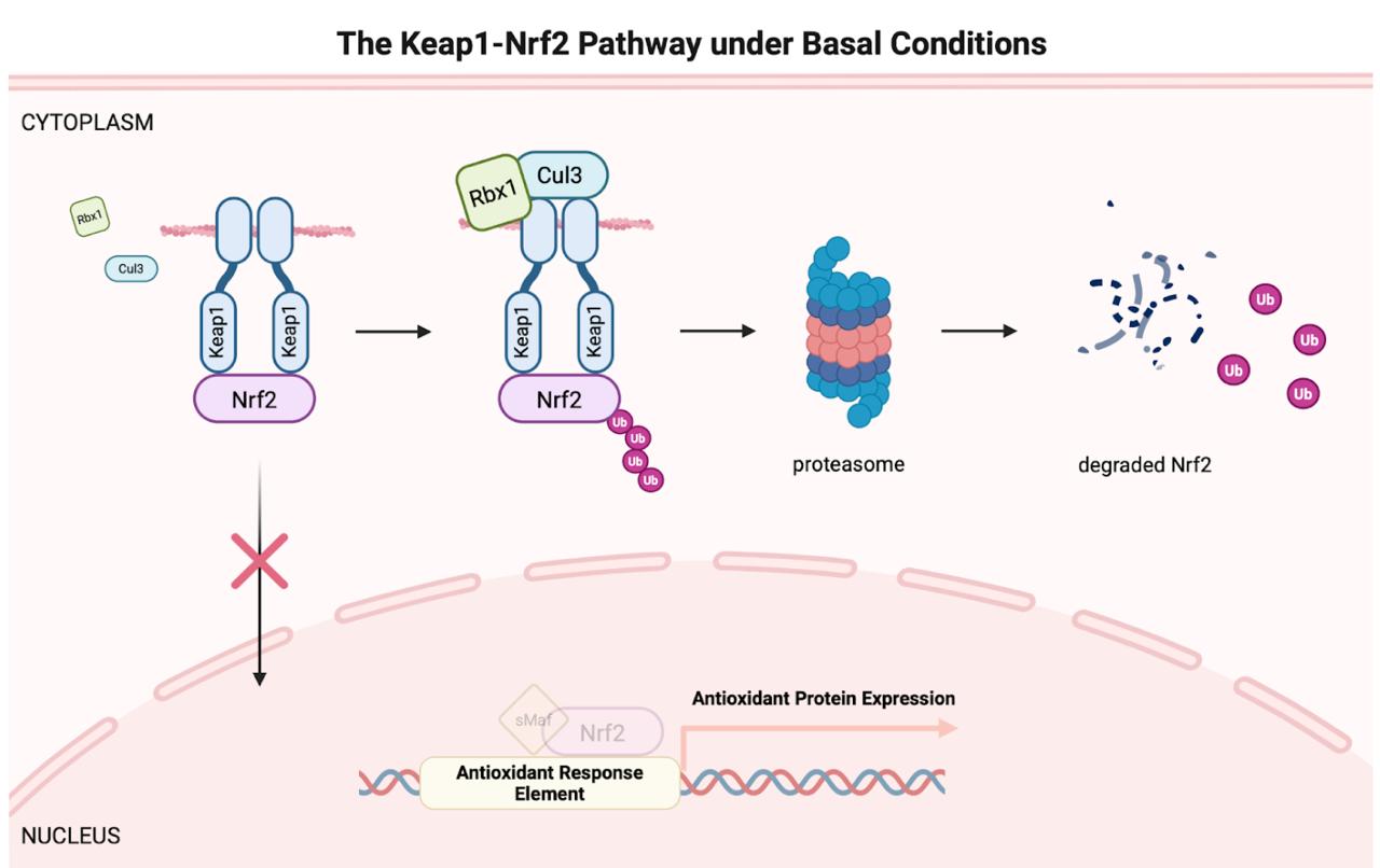
Figure 3. The Keap1-Nrf2 Pathway under Normal Conditions
However, when ROS or electrophilic compounds such as p-benzoquinone accumulate, they react with specific cysteine residues on Keap1. This disrupts Keap1's ability to target Nrf2 for degradation. Without Keap1, Nrf2 becomes free and stabilized. The free Nrf2 then translocates into the nucleus and dimerizes with another protein sMaf to bind together to specific DNA sequences known as Antioxidant Response Elements (AREs) located in the promoters of target genes. These genes encode various antioxidant and detoxifying enzymes, including those involved in quinone detoxification and redox homeostasis (Figure 4).
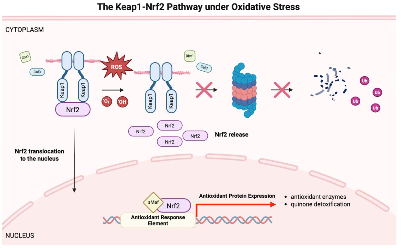
Figure 4. The Keap1-Nrf2 Pathway under Oxidative Stress
ARE-minCMV-EGFP Construct For a Bio-sensor
In our project, we aim to harness this beautiful natural defense mechanism and enhance it with new features that allow rapid detection of hydroquinone exposure. To achieve this, we obtained the Antioxidant Response Element (ARE) and placed it upstream of a minimal CMV promoter. The minimal CMV promoter was chosen specifically because it maintains low basal transcriptional activity and becomes activated only when the ARE is bound by the Nrf2 transcription factor.
When cells experience oxidative stress due to excessive amounts of hydroquinone, this intrinsic defense pathway is immediately triggered. The activated Nrf2 moves into the nucleus and binds to the ARE sequence, initiating transcription of downstream genes. In our design, it is the EGFP reporter gene. Thus, fluorescence expression serves as a visible signal indicating the presence of oxidative stress (Figure 5).
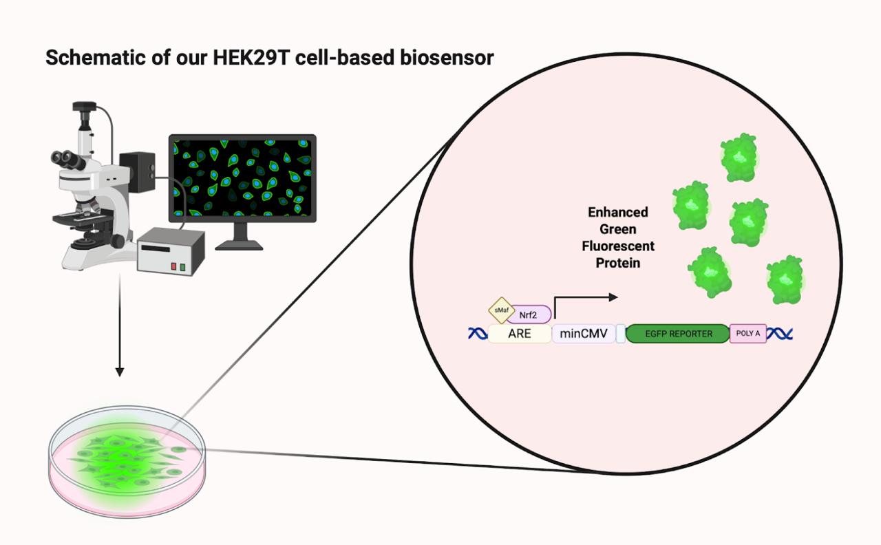
Figure 5. The Proof-of-concept for our bio-sensor based on the ARE-minCMV-EGFP construct
In this proof-of-concept experiment, we aim to demonstrate that HEK293T cells can sense hydroquinone exposure and produce EGFP in response, confirming successful pathway activation. In the future, we plan to expand this system into a dose-dependent biosensor to enable us quantitative detection of oxidative stress levels and improving sensitivity for potential applications in product safety testing and environmental monitoring.
REFERENCES
- Sarkar, R., & Garg, S. (2021). Tips for managing post-inflammatory hyperpigmentation of acne. CosmoDerma, 1, 14. https://cosmoderma.org/tips-for-managing-post-inflammatory-hyperpigmentation-of-acne/
- IARC Working Group on the Evaluation of Carcinogenic Risks to Humans. (1999). Re-evaluation of some organic chemicals, hydrazine and hydrogen peroxide (IARC Monographs, No. 71). Lyon, France: International Agency for Research on Cancer. Section "Hydroquinone." In NCBI Bookshelf. https://www.ncbi.nlm.nih.gov/books/NBK499038/
- Fuertes-Agudo, M., Luque-Tévar, M., Cucarella, C., Martín-Sanz, P., & Casado, M. (2023). Advances in understanding the role of NRF2 in liver pathophysiology and its relationship with hepatic-specific cyclooxygenase-2 expression. Antioxidants, 12, 1491. https://doi.org/10.3390/antiox12081491
- Rushmore, T. H., Morton, M. R., & Pickett, C. B. (1991). The antioxidant responsive element: Activation by oxidative stress and identification of the DNA consensus sequence required for functional activity. Journal of Biological Chemistry, 266, 11632–11639.
- Itoh, K., Chiba, T., Takahashi, S., Ishii, T., Igarashi, K., Katoh, Y., Oyake, T., Hayashi, N., Satoh, K., Hatayama, I., et al. (1997). An Nrf2/Small Maf heterodimer mediates the induction of phase II detoxifying enzyme genes through antioxidant response elements. Biochemical and Biophysical Research Communications, 236, 313–322. https://www.sciencedirect.com/science/article/abs/pii/S0006291X97969436?via%3Dihub
- Itoh, K., Wakabayashi, N., Katoh, Y., Ishii, T., Igarashi, K., Engel, J. D., & Yamamoto, M. (1999). Keap1 represses nuclear activation of antioxidant responsive elements by Nrf2 through binding to the amino-terminal Neh2 domain. Genes & Development, 13, 76–86.
- Moi, P., Chan, K., Asunis, I., Cao, A., & Kan, Y. W. (1994). Isolation of NF-E2-related factor 2 (Nrf2), a NF-E2-like basic leucine zipper transcriptional activator that binds to the tandem NF-E2/AP1 repeat of the beta-globin locus control region. Proceedings of the National Academy of Sciences of the United States of America, 91, 9926–9930. https://www.pnas.org/doi/abs/10.1073/pnas.91.21.9926